Certain cephalopods like cuttlefish, octopuses, and squid have the ability to camouflage themselves by making themselves transparent and/or changing their coloration. Scientists would like to learn more about the precise mechanisms underlying this unique ability, but it's not possible to culture squid skin cells in the lab. Researchers at the University of California, Irvine, have discovered a viable solution: replicating the properties of squid skin cells in mammalian (human) cells in the lab. They presented their research at a meeting of the American Chemical Society being held this week in Indianapolis.
"In general, there's two ways you can achieve transparency," UC Irvine's Alon Gorodetsky, who has been fascinated by squid camouflage for the last decade or so, said during a media briefing at the ACS meeting. "One way is by reducing how much light is absorbed—pigment-based coloration, typically. Another way is by changing how light is scattered, typically by modifying differences in the refractive index." The latter is the focus of his lab's research.
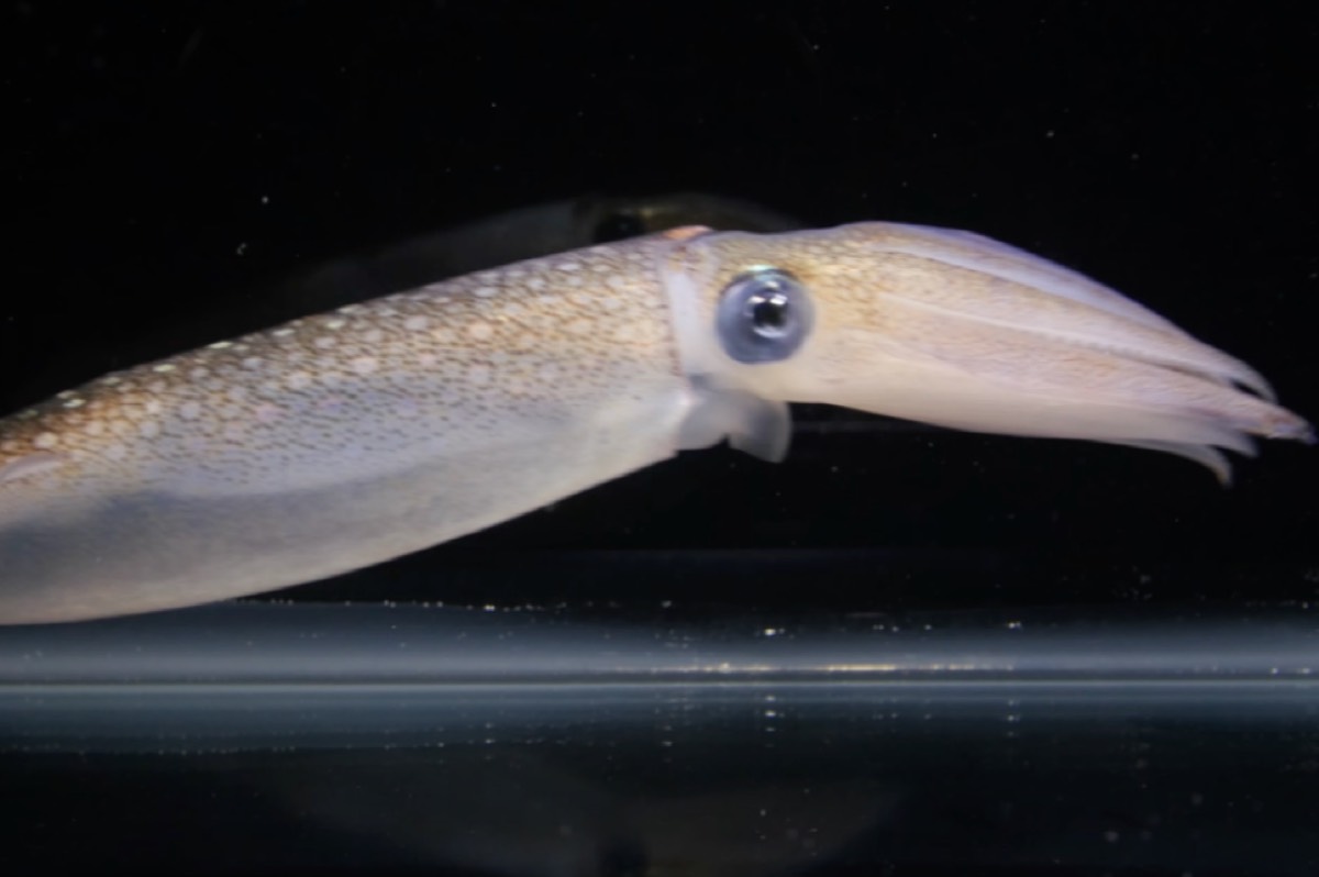
Squid skin is translucent and features an outer layer of pigment cells called chromatophores that control light absorption. Each chromatophore is attached to muscle fibers that line the skin's surface, and those fibers, in turn, are connected to a nerve fiber. It's a simple matter to stimulate those nerves with electrical pulses, causing the muscles to contract. And because the muscles pull in different directions, the cell expands, along with the pigmented areas, which changes the color. When the cell shrinks, so do the pigmented areas.
Underneath the chromatophores, there is a separate layer of iridophores. Unlike the chromatophores, the iridophores aren't pigment-based but are an example of structural color, similar to the crystals in the wings of a butterfly, except a squid's iridophores are dynamic rather than static. They can be tuned to reflect different wavelengths of light. A 2012 paper suggested that this dynamically tunable structural color of the iridophores is linked to a neurotransmitter called acetylcholine. The two layers work together to generate the unique optical properties of squid skin.
And then there are leucophores, similar to the iridophores except they scatter the full spectrum of light, such that they appear white. They contain reflectin proteins that typically clump together into nanoparticles, so that light scatters instead of being absorbed or directly transmitted. Leucophores are mostly found in cuttlefish and octopuses, but there are some female squid of the genus Sepioteuthis that have leucophores that they can 'tune" to only scatter certain wavelengths of light. If the cells allow light through with little scattering, they’ll seem more transparent, while the cells become opaque and more apparent by scattering a lot more light. These are the cells that interest Gorodetsky.
In 2015, Gorodetsky's lab created squid-inspired invisibility stickers to one day help soldiers disguise themselves, even from infrared cameras. The stickers were essentially thin, flexible layers of camo with the potential to take on a pattern to match the soldiers’ infrared reflectance to their background. Rather than killing squid to collect the reflectin proteins, they could express it in e. coli bacterial cultures. Then they coated the equivalent of common household packing tape with the modified bacteria. The stickers could be tuned merely by changing the thickness of the bacterial film. Thinner films appeared blue; thicker films appeared orange.
Having already experimented with truncated versions of the protein to study its refractive index and how it scatters light, Gorodetsky's team has now extended that research by introducing squid-derived genes that encode reflectin into human cells. The trick was to get reflectin nanostructures to form stably rather than temporarily. Adding salt to the cells' culture media caused the reflectin to clump together into light-scattering nanoparticles, and by gradually increasing the salt concentrations, the nanoparticles became larger so that even more light was scattered, essentially "tuning" their opacity. They took detailed time-lapse images of the nanoparticles' properties using a technique called holotomography.
"We were really trying to understand whether the intrinsic characteristics of these proteins—their high refractive indices, their ability to self-assemble into specific structures—could be replicated in a mammalian cell," said Gorodetsky. "So we engineered mammalian cells to express large amounts of this protein. And we found that the [resulting] self-assembled structures were very similar in many ways in terms of their sizes and their optical properties."
When the COVID-19 pandemic hit, and it wasn't possible to work in the lab, Gorodetsky's graduate student, Georgii Bogdanov, used the imaging data to create a computational model, enabling them to make predictions and compare the optical properties of squid cells and their engineered mammalian cells. "The refractive indices are comparable, which is the key component of this phenomenon," said Bogdanov. "And while the sizes of those particles are also similar, this gives a perfect comparison of the light scattering that happens in the squid skin and mammalian cells."
What about potential applications? Earlier this year, we reported that engineers at the University of Toronto drew inspiration from the squid to create a prototype for "liquid windows" that can shift the wavelength, intensity, and distribution of light transmitted through those windows, thereby saving substantially on energy costs. Gorodetsky said that one potential application of his own research is using reflectin proteins as sub-cellular molecular probes with a high refractive index, used in conjunction with advanced microscopy techniques. Such genetically encoded tags would not bleach inside human cells, enabling scientists to track cell structure to gain a better understanding of cell growth and development.
DOI: ACS Biomaterials Science & Engineering, 2023. 10.1021/acsbiomaterials.2c00088 (About DOIs).